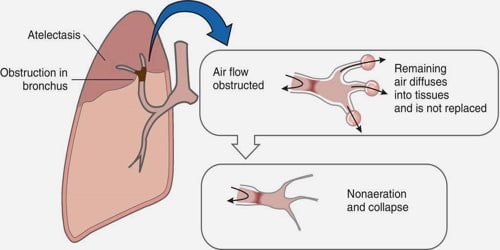
Anaesthesia and bronchoscopy are two of the most common methods for diagnosing atelectasis. Bronchoscopy involves the use of a long tube called a bronchoscope to view the lungs and the airways through a camera. A tracheoscope is used to examine the chest and neck for obstructions and abnormalities. For patients who have a history of severe asthma, bronchoscopy may not be feasible due to their risk of aspiration pneumonia.
Aortic stenosis, the narrowing or blocking of an air passage (bronchi or bronchioles), or by increased pressure on the respiratory organ, is the primary cause of atelectasis. Common risk factors for atelectasis are prolonged bed rest with little change in position and airway obstruction due to a decreased alveolar ventilation. Other potential causes of atelectasis may include viral infections, hyperthyroidism and pregnancy. Patients with atelectasis have been diagnosed as having obstructive sleep apnea and can suffer from pulmonary embolism, resulting in cardiac arrest and death. Patients with sleep apnea are at increased risk for atelectasis.
In cases of chronic obstructive pulmonary disorder (COPD), which is often associated with sleep apnea, patients are at a higher risk for developing atelectasis. COPD is a disease that affects the respiratory system and can lead to serious complications such as congestive heart failure. Patients with COPD are at a greater risk of developing atelectasis because their lungs are unable to effectively exchange carbon dioxide for oxygen.
The diagnosis of atelectasis is made based on the symptoms present, including wheezing, choking, breathing that has difficulty in locating the bronchial tubes, coughing, shortness of breath, chest pain and chest discomfort, and frequent episodes of asthma. A thorough physical exam will need to be performed to exclude other conditions. If atelectasis is present, a bronchoscope can be used to view the lungs and breathing passages through a camera.
There are several surgical procedures available for the treatment of atelectasis. The most common surgical procedure is referred to as "surgical perforation". This involves the insertion of a balloon catheter into the airway to provide access to the lungs for the removal of obstructions. or the replacement of airways.
Surgical perforation of the atelectasis is an invasive procedure that requires incisions to be made in the chest. It is done under general anesthesia. This procedure is often followed by the insertion of a drainage cannula to collect the collected fluid and the placement of a catheter to collect it.

Once the catheter is in place, the drainage cannula can be used to remove the fluid through tubing into a collection bag
During the recovery period, patients are advised to take antihistamines for any possible side effects and to avoid exertion. Patients are allowed to take small amounts of medication if symptoms become worse. After the surgery, patients are advised to keep the incisions clean and free of blood.
Patients who have had this surgery should stay in the hospital for several days to allow the incision to heal. They are given pain medication and monitored closely to prevent infection. Antibiotics may be given to prevent infection and promote healing.
Surgical perforation of the atelectasis is effective in treating most cases, but it is not without risks. Infection can occur anywhere the catheter enters the lung or through the draining catheter and can lead to a condition known as disseminated intravascular coagulation. This occurs when the catheter is located outside the lungs where the spread of infection can occur.
There are several methods used to remove catheter drainage from the lungs and to drain the fluid collected. The most commonly used technique is the mechanical drainage method. This is done by a bag-valve pump that sucks out the fluid by creating suction with the help of a tube.
This method is less invasive and does not require the insertion of a catheter. A catheter is used instead and is placed just outside the lungs, where it provides the suction needed for the drainage process.
In addition, the patient may be advised to wear a mask or use a special breathing device while they recover. This allows for improved oxygenation and the possibility of infection is minimal.
